




Specialist Surgery for Parotid Gland Tumours
Parotid gland tumours arise within the largest of the salivary glands, located just in front of and below the ear.
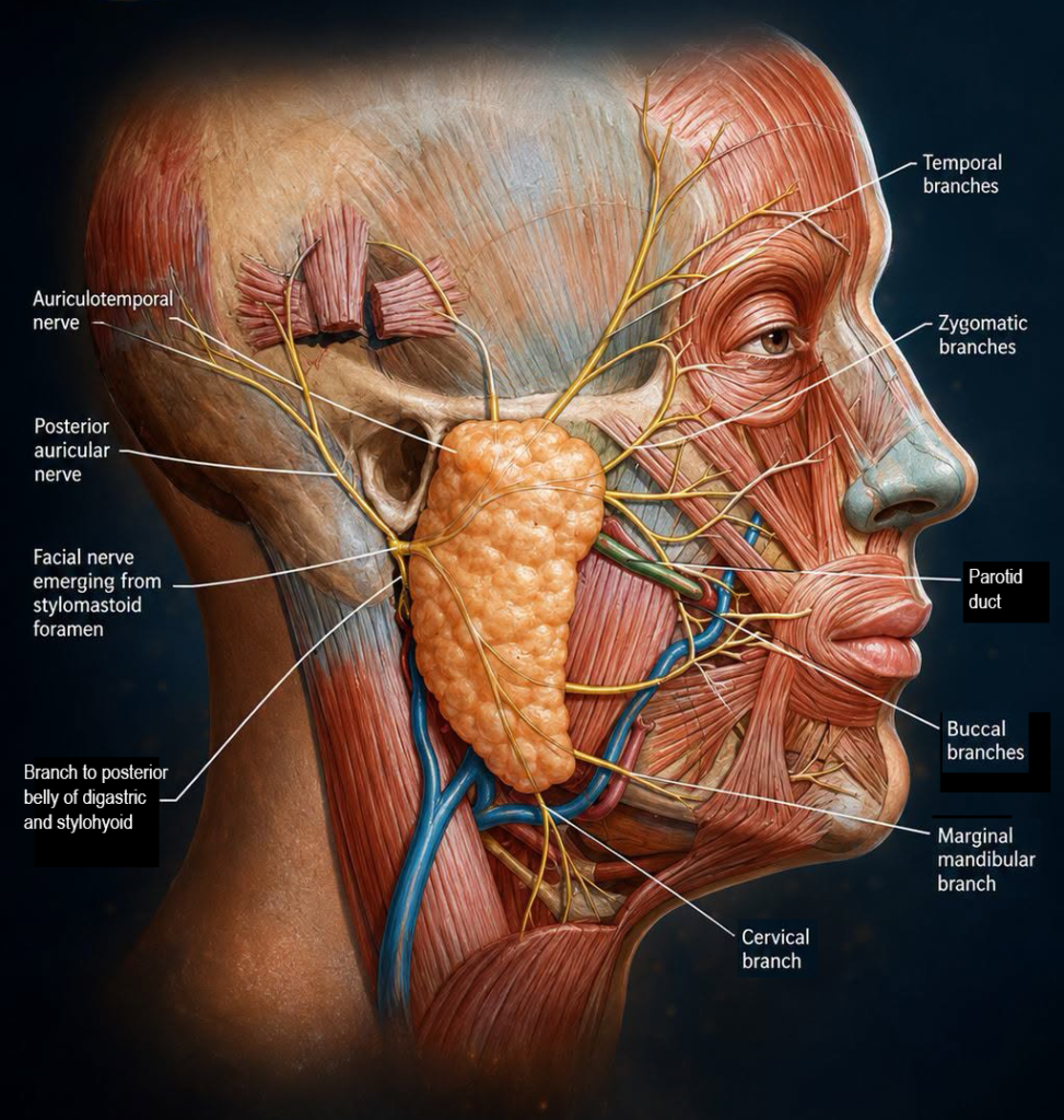
Surgical removal is the primary treatment for most parotid tumours. The procedure requires careful planning due to the presence of the facial nerve, which runs through the gland and controls facial movement.
The aim is to remove the tumour safely while preserving nerve function and achieving a balanced cosmetic outcome.
Understanding Parotid Tumours
Parotid tumours may be:
- Benign (most common, e.g. pleomorphic adenoma, Warthin’s tumour)
- Malignant (less common, but require more extensive treatment)
Even benign tumours are usually removed, as they can:
- Continue to grow over time
- Become more complex to treat
- Occasionally undergo change
The Role of Surgery
Surgery aims to:
- Remove the tumour and send it for histological analysis
- Preserve the facial nerve and its branches
- Minimise the risk of recurrence
- Achieve a well-healed, discreet scar
The exact technique depends on the size, location, and nature of the tumour.
Surgical Techniques
Partial Parotidectomy
This procedure involves removal of the portion of the gland containing the tumour together with careful identification and dissection of the facial nerve.
Advantages include:
- A reliable and well-established operation with excellent long-term outcomes
- Suitable for a wider range of tumours and clinical situations
- Direct visualisation of facial nerve branches
- Access to deeper tumours
Extracapsular Dissection
ECD involves removing the tumour together with a thin margin of surrounding healthy tissue, without routinely exposing the main trunk of the facial nerve.
Compared with traditional parotid surgery, ECD offers several potential advantages:
- Less disruption of normal parotid tissue
- Reduced surgical dissection
- Reduced risk of contour deformity (“hollowing”) around the jaw and cheek
- Faster recovery
Importantly, ECD is not suitable for every tumour. Careful patient selection using imaging, biopsy results, tumour size and tumour location is essential.
When used appropriately, modern studies have shown ECD to provide excellent tumour control with low recurrence rates and outcomes comparable to more extensive procedures for selected benign tumours.
Facelift Incision (Hidden Scar Parotid Surgery)
Whenever appropriate, I use a hidden facelift incision rather than the traditional, more noticeable modified Blair parotidectomy incision.
The incision is concealed within the natural contours around the ear and extends into the hairline, allowing access to the parotid gland whilst minimising visible scarring.
The potential benefits of a facelift incision for parotid surgery include avoidance of a visible neck scar, and hence improved cosmetic outcome.
The facelift approach can be used in conjunction with either extracapsular dissection or partial parotidectomy, as it relates to the incision design rather than the extent of glad removal.
Many patients are surprised that major salivary gland surgery can often be performed through an incision that becomes difficult to detect once fully healed.
Facial Nerve Preservation and Monitoring
The facial nerve is central to parotid surgery, controlling movement of the face.
During surgery:
- The nerve and its branches are carefully identified and preserved
- Intraoperative facial nerve monitoring is used to assist this process
Facial nerve monitoring works by:
- Providing real-time feedback on nerve function
- Helping identify nerve branches during dissection
- Reducing the risk of inadvertent injury
This adds an additional level of safety, particularly in:
- More complex tumours
- Revision surgery
- Cases where anatomy is distorted
In the majority of cases, normal facial movement is preserved. Temporary weakness can occur and usually improves over time.
Recovery
Recovery depends on the extent of surgery.
- A short hospital stay is usually required
- A small surgical drain may present for the first few days
- Swelling and bruising settle over several weeks
- Return to normal activity is gradual
Follow-up ensures appropriate healing and functional recovery.
Risks and Considerations
Potential risks include:
- Temporary or, rarely, permanent facial nerve weakness
- Numbness around the ear
- Bleeding or infection (uncommon)
- Salivary leak or fluid collection
- Frey’s syndrome (sweating when eating)
- Recurrence depending on tumour type
Careful planning, technique, and nerve monitoring help minimise these risks.
Consultation
A detailed consultation includes assessment of the lump, imaging where required, and discussion of the most appropriate surgical approach.
Book a Consultation
For assessment and treatment of parotid gland tumours in London, arrange a consultation to discuss your case in detail.